AMIC® Chondro-Gide® im Talus
Chondrale und osteochondrale Läsionen (OCL) des Sprunggelenks kommen häufig vor und werden zunehmend als Ursache für anhaltende Schmerzen im Sprunggelenk erkannt. Die meisten osteochondralen Läsionen des Talus (OLT) sind auf Traumata und wiederholte Mikrotraumata1 zurückzuführen. Etwa 50 % der Knöchelverstauchungen und bis zu 73 % der Sprunggelenksfrakturen führen zu einer Knorpelverletzung und können entsprechende Symptome auslösen.2
Knochenmarkstimulation empfohlen für OCL <1 cm2
Ramponi et. al. schlugen vor, Knochenmarkstimulationstechniken auf OCL < 1 cm2 zu beschränken. Der Grenzwert von 1 cm2 wurde 2017 auch in einem Konsensmeeting der International Society on Cartilage Repair of the Ankle (ISCRA) bestätigt. Diese und weitere Konsensstatements finden Sie unter https://my.on-foundation.org/focus.
ISCRA legte die idealen Grössen für Knochenmarkstimulation wie folgt fest: Durchmesser < 10 mm, Bereich < 100 mm2, Tiefe < 5 mm.4 Ab einer Tiefe von > 3 mm kann bereits ein Knochenaufbau in Erwägung gezogen werden. Laut aktueller Literatur unterstützt die Konsensusempfehlung die Verwendung eines Scaffolds für den Knochenaufbau.
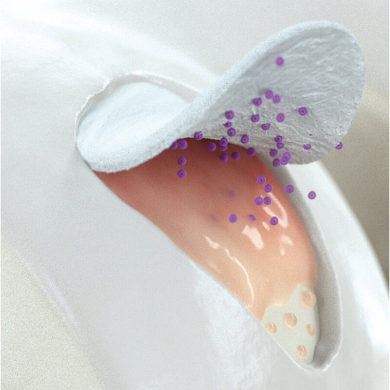
AMIC® Chondro-Gide® für wirksame Knorpelreparatur
AMIC® Chondro-Gide® ist ein minimalinvasives einzeitiges Verfahren, das Knochenmarkstimulation in Kombination mit Chondro-Gide® nutzt, um Knorpeldefekte jeder Grösse zu reparieren. Es kann entweder durch Osteotomie6 oder Mini-open-Chirurgie7 durchgeführt werden.
AMIC® Chondro-Gide® wurde von Geistlich Surgery in Zusammenarbeit mit führenden europäischen Chirurgen entwickelt und ist eine effektive und kostengünstige Behandlung7 zur Reparatur von Knorpelläsionen im Talus, zur Schmerzprophylaxe und -linderung sowie zur Verlangsamung des Knorpelabbaus.
References
- CHEW, K. T. L., 2008, Osteochondral lesions of the talus. Annals of the Academy of Medicine. 2008. Vol.37, no. 1, p. 63-8
- STEELE, J. R., et al., Osteochondral Lesions of the Talus. Foot & Ankle Orthopaedics. 2018. Vol. 3, no. 3, p. 247301141877955. DOI 10.1177/2473011418779559. SAGE Publications
- RAMPONI, L., et al., Lesion Size Is a Predictor of Clinical Outcomes After Bone Marrow Stimulation for Osteochondral Lesions of the Talus: A Systematic Review. The American Journal of Sports Medicine. 2016. Vol. 45, no. 7, p. 1698-1705. DOI10.1177/0363546516668292. SAGE Publications (Systematic Review)
- HANNON et al. Debridement, Curettage, Microfracture, and Fixation Techniques for Osteochondral Lesions of the Talus, 2018. Foot & Ankle Orthopaedics, Vol. 3, no. 3, p. 2473011418S0006. DOI 10.1177/2473011418s00066. SAGE Publications (Consensus Meeting Report)
- ROTHRAUFF, B.B., et al., Scaffold-Based Therapies: Proceedings of the International Consensus Meeting on Cartilage Repair of the Ankle. Foot & Ankle International. 2018. Vol. 39, no. 1_suppl, p. 41S-47S. DOI 10.1177/1071100718781864. SAGE Publications (Consensus Meeting)24. WALTHER, M., ALTENBERGER, S., KRIEGELSTEIN, S., VOLKERING, C. and R.SER, A., 2014, Reconstruction of focal cartilage defects in the talus with miniarthrotomy and collagen matrix. Operative Orthop.die und Traumatologie. 2014. Vol. 26, no. 6, p. 603-610. DOI 10.1007/s00064-012-0229-9. Springer Nature (Clinical Study)
- VALDERRABANO, V., et al., Reconstruction of Osteochondral Lesions of the Talus With Autologous Spongiosa Grafts and Autologous Matrix-Induced Chondrogenesis. The American Journal of Sports Medicine. 2013. Vol. 41, no. 3, p. 519-527.DOI 10.1177/0363546513476671. SAGE Publications (Clinical Study)
- WALTHER, M., et al., Reconstruction of focal cartilage defects in the talus with miniarthrotomy and collagen matrix. Operative Orthop.die und Traumatologie. 2014. Vol. 26, no. 6, p. 603-610. DOI 10.1007/s00064-012-0229-9. Springer Nature (Clinical Study)
- GOTTSCHALK, O., et al., Functional Medium-Term Results After Autologous Matrix-Induced Chondrogenesis for Osteochondral Lesions of the Talus: A 5-Year Prospective Cohort Study. The Journalof Foot and Ankle Surgery. 2017. Vol. 56, no. 5, p. 930-936. DOI 10.1053/j.jfas.2017.05.002. Elsevier BV (Clinical Study)
- YOUNG, KI WON, et al., Medial approaches to osteochondral lesion of the talus without medial malleolar osteotomy. Knee Surgery, Sports Traumatology, Arthroscopy. 2009. Vol. 18, no. 5, p. 634-637. DOI 10.1007/s00167-009-1019-2. Springer Nature
- GALLA, MELLANY, DUENSING, IAN, KAHN, TIMOTHY L. and BARG, ALEXEJ, 2018, Open reconstruction with autologous spongiosa grafts and matrix-induced chondrogenesis for osteochondral lesions of the talus can be performed without medial malleolar osteotomy. Knee Surgery, Sports Traumatology, Arthroscopy. 2018. DOI 10.1007/s00167-018-5063-7. Springer Nature (Clinical Study)
- Chondro-Gide® IFU 2019, Geistlich Pharma AG
- WALTHER, M., et al., Reconstruction of focal cartilage defects in the talus with miniarthrotomy and collagen matrix. Operative Orthopädie und Traumatologie. 2014. Vol. 26, no. 6, p. 603-610. DOI 10.1007/s00064-012-0229-9. Springer Nature (Clinical Study)
- GOTTSCHALK, O., et al., Functional Medium-Term Results After Autologous Matrix-Induced Chondrogenesis for Osteochondral Lesions of the Talus: A 5-Year Prospective Cohort Study. The Journal of Foot and Ankle Surgery. 2017. Vol. 56, no. 5, p. 930-936. DOI 10.1053/j.jfas.2017.05.002. Elsevier BV (Clinical Study)
- USUELLI, F., et al., All-arthroscopic AMIC® (AT-AMIC®) technique with autologous bone graft for talar osteochondral defects: clinical and radiological results. Knee Surgery, Sports Traumatology, Arthroscopy. 2016. Vol. 26, no. 3, p. 875-881. DOI 10.1007/s00167-016-4318-4. Springer Nature (Clinical Study)


Referenzen
- CHEW, K. T. L., 2008, Osteochondral lesions of the talus. Annals of the Academy of Medicine. 2008. Vol.37, no. 1, p. 63-8
- STEELE, J. R., et al., Osteochondral Lesions of the Talus. Foot & Ankle Orthopaedics. 2018. Vol. 3, no. 3, p. 247301141877955. DOI 10.1177/2473011418779559. SAGE Publications
- RAMPONI, L., et al., Lesion Size Is a Predictor of Clinical Outcomes After Bone Marrow Stimulation for Osteochondral Lesions of the Talus: A Systematic Review. The American Journal of Sports Medicine. 2016. Vol. 45, no. 7, p. 1698-1705. DOI10.1177/0363546516668292. SAGE Publications (Systematic Review)
- HANNON et al. Debridement, Curettage, Microfracture, and Fixation Techniques for Osteochondral Lesions of the Talus, 2018. Foot & Ankle Orthopaedics, Vol. 3, no. 3, p. 2473011418S0006. DOI 10.1177/2473011418s00066. SAGE Publications (Consensus Meeting Report)
- ROTHRAUFF, B.B., et al., Scaffold-Based Therapies: Proceedings of the International Consensus Meeting on Cartilage Repair of the Ankle. Foot & Ankle International. 2018. Vol. 39, no. 1_suppl, p. 41S-47S. DOI 10.1177/1071100718781864. SAGE Publications (Consensus Meeting)24. WALTHER, M., ALTENBERGER, S., KRIEGELSTEIN, S., VOLKERING, C. and R.SER, A., 2014, Reconstruction of focal cartilage defects in the talus with miniarthrotomy and collagen matrix. Operative Orthop.die und Traumatologie. 2014. Vol. 26, no. 6, p. 603-610. DOI 10.1007/s00064-012-0229-9. Springer Nature (Clinical Study)
- VALDERRABANO, V., et al., Reconstruction of Osteochondral Lesions of the Talus With Autologous Spongiosa Grafts and Autologous Matrix-Induced Chondrogenesis. The American Journal of Sports Medicine. 2013. Vol. 41, no. 3, p. 519-527.DOI 10.1177/0363546513476671. SAGE Publications (Clinical Study)
- WALTHER, M., et al., Reconstruction of focal cartilage defects in the talus with miniarthrotomy and collagen matrix. Operative Orthop.die und Traumatologie. 2014. Vol. 26, no. 6, p. 603-610. DOI 10.1007/s00064-012-0229-9. Springer Nature (Clinical Study)
- GOTTSCHALK, O., et al., Functional Medium-Term Results After Autologous Matrix-Induced Chondrogenesis for Osteochondral Lesions of the Talus: A 5-Year Prospective Cohort Study. The Journalof Foot and Ankle Surgery. 2017. Vol. 56, no. 5, p. 930-936. DOI 10.1053/j.jfas.2017.05.002. Elsevier BV (Clinical Study)
- YOUNG, KI WON, et al., Medial approaches to osteochondral lesion of the talus without medial malleolar osteotomy. Knee Surgery, Sports Traumatology, Arthroscopy. 2009. Vol. 18, no. 5, p. 634-637. DOI 10.1007/s00167-009-1019-2. Springer Nature
- GALLA, MELLANY, DUENSING, IAN, KAHN, TIMOTHY L. and BARG, ALEXEJ, 2018, Open reconstruction with autologous spongiosa grafts and matrix-induced chondrogenesis for osteochondral lesions of the talus can be performed without medial malleolar osteotomy. Knee Surgery, Sports Traumatology, Arthroscopy. 2018. DOI 10.1007/s00167-018-5063-7. Springer Nature (Clinical Study)
- Chondro-Gide® IFU 2019, Geistlich Pharma AG
- WALTHER, M., et al., Reconstruction of focal cartilage defects in the talus with miniarthrotomy and collagen matrix. Operative Orthopädie und Traumatologie. 2014. Vol. 26, no. 6, p. 603-610. DOI 10.1007/s00064-012-0229-9. Springer Nature (Clinical Study)
- GOTTSCHALK, O., et al., Functional Medium-Term Results After Autologous Matrix-Induced Chondrogenesis for Osteochondral Lesions of the Talus: A 5-Year Prospective Cohort Study. The Journal of Foot and Ankle Surgery. 2017. Vol. 56, no. 5, p. 930-936. DOI 10.1053/j.jfas.2017.05.002. Elsevier BV (Clinical Study)
- USUELLI, F., et al., All-arthroscopic AMIC® (AT-AMIC®) technique with autologous bone graft for talar osteochondral defects: clinical and radiological results. Knee Surgery, Sports Traumatology, Arthroscopy. 2016. Vol. 26, no. 3, p. 875-881. DOI 10.1007/s00167-016-4318-4. Springer Nature (Clinical Study)
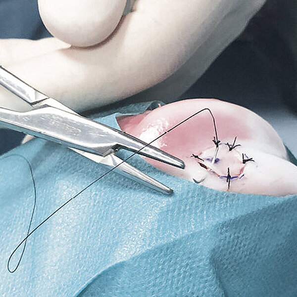
Mini - Open Technik nach Prof. Dr. M. Walther


Referenzen
- CHEW, K. T. L., 2008, Osteochondral lesions of the talus. Annals of the Academy of Medicine. 2008. Vol.37, no. 1, p. 63-8
- STEELE, J. R., et al., Osteochondral Lesions of the Talus. Foot & Ankle Orthopaedics. 2018. Vol. 3, no. 3, p. 247301141877955. DOI 10.1177/2473011418779559. SAGE Publications
- RAMPONI, L., et al., Lesion Size Is a Predictor of Clinical Outcomes After Bone Marrow Stimulation for Osteochondral Lesions of the Talus: A Systematic Review. The American Journal of Sports Medicine. 2016. Vol. 45, no. 7, p. 1698-1705. DOI10.1177/0363546516668292. SAGE Publications (Systematic Review)
- HANNON et al. Debridement, Curettage, Microfracture, and Fixation Techniques for Osteochondral Lesions of the Talus, 2018. Foot & Ankle Orthopaedics, Vol. 3, no. 3, p. 2473011418S0006. DOI 10.1177/2473011418s00066. SAGE Publications (Consensus Meeting Report)
- ROTHRAUFF, B.B., et al., Scaffold-Based Therapies: Proceedings of the International Consensus Meeting on Cartilage Repair of the Ankle. Foot & Ankle International. 2018. Vol. 39, no. 1_suppl, p. 41S-47S. DOI 10.1177/1071100718781864. SAGE Publications (Consensus Meeting)24. WALTHER, M., ALTENBERGER, S., KRIEGELSTEIN, S., VOLKERING, C. and R.SER, A., 2014, Reconstruction of focal cartilage defects in the talus with miniarthrotomy and collagen matrix. Operative Orthop.die und Traumatologie. 2014. Vol. 26, no. 6, p. 603-610. DOI 10.1007/s00064-012-0229-9. Springer Nature (Clinical Study)
- VALDERRABANO, V., et al., Reconstruction of Osteochondral Lesions of the Talus With Autologous Spongiosa Grafts and Autologous Matrix-Induced Chondrogenesis. The American Journal of Sports Medicine. 2013. Vol. 41, no. 3, p. 519-527.DOI 10.1177/0363546513476671. SAGE Publications (Clinical Study)
- WALTHER, M., et al., Reconstruction of focal cartilage defects in the talus with miniarthrotomy and collagen matrix. Operative Orthop.die und Traumatologie. 2014. Vol. 26, no. 6, p. 603-610. DOI 10.1007/s00064-012-0229-9. Springer Nature (Clinical Study)
- GOTTSCHALK, O., et al., Functional Medium-Term Results After Autologous Matrix-Induced Chondrogenesis for Osteochondral Lesions of the Talus: A 5-Year Prospective Cohort Study. The Journalof Foot and Ankle Surgery. 2017. Vol. 56, no. 5, p. 930-936. DOI 10.1053/j.jfas.2017.05.002. Elsevier BV (Clinical Study)
- YOUNG, KI WON, et al., Medial approaches to osteochondral lesion of the talus without medial malleolar osteotomy. Knee Surgery, Sports Traumatology, Arthroscopy. 2009. Vol. 18, no. 5, p. 634-637. DOI 10.1007/s00167-009-1019-2. Springer Nature
- GALLA, MELLANY, DUENSING, IAN, KAHN, TIMOTHY L. and BARG, ALEXEJ, 2018, Open reconstruction with autologous spongiosa grafts and matrix-induced chondrogenesis for osteochondral lesions of the talus can be performed without medial malleolar osteotomy. Knee Surgery, Sports Traumatology, Arthroscopy. 2018. DOI 10.1007/s00167-018-5063-7. Springer Nature (Clinical Study)
- Chondro-Gide® IFU 2019, Geistlich Pharma AG
- WALTHER, M., et al., Reconstruction of focal cartilage defects in the talus with miniarthrotomy and collagen matrix. Operative Orthopädie und Traumatologie. 2014. Vol. 26, no. 6, p. 603-610. DOI 10.1007/s00064-012-0229-9. Springer Nature (Clinical Study)
- GOTTSCHALK, O., et al., Functional Medium-Term Results After Autologous Matrix-Induced Chondrogenesis for Osteochondral Lesions of the Talus: A 5-Year Prospective Cohort Study. The Journal of Foot and Ankle Surgery. 2017. Vol. 56, no. 5, p. 930-936. DOI 10.1053/j.jfas.2017.05.002. Elsevier BV (Clinical Study)
- USUELLI, F., et al., All-arthroscopic AMIC® (AT-AMIC®) technique with autologous bone graft for talar osteochondral defects: clinical and radiological results. Knee Surgery, Sports Traumatology, Arthroscopy. 2016. Vol. 26, no. 3, p. 875-881. DOI 10.1007/s00167-016-4318-4. Springer Nature (Clinical Study)
Klinische Ergebnisse AMIC® Chondro-Gide® Talus
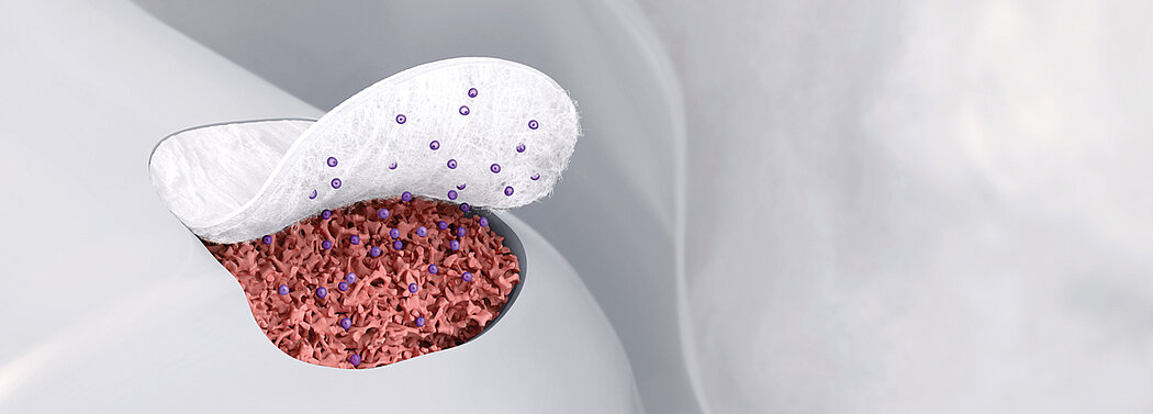
Wie Chondro-Gide® wirkt
Chondro-Gide® bildet eine Schutzschicht, die dafür sorgt, dass die aus dem Knochen freigesetzten oder in den Defekt eingeführten Zellen an Ort und Stelle bleiben. Die Membran unterstützt die regenerative Behandlung von chondralen und osteochondralen Läsionen. In der Anfangsphase des Heilungsprozesses werden der Defekt und die Zellen zunächst mit Chondro-Gide® abgedeckt. Nach etwa 1-4 Monaten11 wird die Matrix dann resorbiert und durch körpereigenes Gewebe ersetzt.
Chondro-Gide® ohne Osteotomie erfolgreich
In mehreren Studien wurde gezeigt, dass Chondro-Gide® ohne Osteotomie erfolgreich platziert werden kann.
Walther et al.12 beschrieben die Rekonstruktion fokaler Knorpeldefekte des Talus mittels Miniarthrotomie und Chondro-Gide® für fokale Knorpeldefekte vom ICRS-Grad III und IV mit einer Fläche von > 1,5 cm2. Die Wissenschaftler überprüften die postoperativen Ergebnisse von 14 Patienten über einen Nachbeobachtungszeitraum von > 30 Monaten. In allen Fällen verbesserten sich die AOFAS-Scores (American Orthopedic Foot and Ankle Society) – zwischen 50 und 89 Punkte –, wobei beide Seiten des oberen Sprunggelenks gleich beweglich waren. Es wurden keine intraoperativen Komplikationen beobachtet. Sie kamen zum Ergebnis, dass es sich bei AMIC® Chondro-Gide® um ein einfaches Verfahren handelt. Die Membran kann in nahezu allen Fällen im Rahmen einer Miniarthrotomie ohne Osteotomie des Schienbein- oder Aussenknöchels implantiert werden. Das einzeitige Verfahren AMIC® Chondro-Gide® wurde darüber hinaus im Vergleich zur Knorpelrekonstruktion mit in vitro gezüchteten Chondrozyten für kosteneffizient befunden.
5-Jahres-Daten
In einer Zwischenanalyse stellten Gottschalk et al.13 vielversprechende Ergebnisse sowohl in der 2-Jahres- als auch in der 5-Jahres-Studie der Patientenkohorte fest.
Die Forscher fanden die größte Verbesserung der klinischen Ergebnisse im ersten Jahr nach der Operation. Zwischen dem 1- und 5-Jahres-Follow-up konnte eine weitere, jedoch statistisch nicht signifikante Verbesserung beobachtet werden. Bezeichnenderweise kehrten die Patienten nach 5 Jahren zum Sport zurück.
References
- CHEW, K. T. L., 2008, Osteochondral lesions of the talus. Annals of the Academy of Medicine. 2008. Vol.37, no. 1, p. 63-8
- STEELE, J. R., et al., Osteochondral Lesions of the Talus. Foot & Ankle Orthopaedics. 2018. Vol. 3, no. 3, p. 247301141877955. DOI 10.1177/2473011418779559. SAGE Publications
- RAMPONI, L., et al., Lesion Size Is a Predictor of Clinical Outcomes After Bone Marrow Stimulation for Osteochondral Lesions of the Talus: A Systematic Review. The American Journal of Sports Medicine. 2016. Vol. 45, no. 7, p. 1698-1705. DOI10.1177/0363546516668292. SAGE Publications (Systematic Review)
- HANNON et al. Debridement, Curettage, Microfracture, and Fixation Techniques for Osteochondral Lesions of the Talus, 2018. Foot & Ankle Orthopaedics, Vol. 3, no. 3, p. 2473011418S0006. DOI 10.1177/2473011418s00066. SAGE Publications (Consensus Meeting Report)
- ROTHRAUFF, B.B., et al., Scaffold-Based Therapies: Proceedings of the International Consensus Meeting on Cartilage Repair of the Ankle. Foot & Ankle International. 2018. Vol. 39, no. 1_suppl, p. 41S-47S. DOI 10.1177/1071100718781864. SAGE Publications (Consensus Meeting)24. WALTHER, M., ALTENBERGER, S., KRIEGELSTEIN, S., VOLKERING, C. and R.SER, A., 2014, Reconstruction of focal cartilage defects in the talus with miniarthrotomy and collagen matrix. Operative Orthop.die und Traumatologie. 2014. Vol. 26, no. 6, p. 603-610. DOI 10.1007/s00064-012-0229-9. Springer Nature (Clinical Study)
- VALDERRABANO, V., et al., Reconstruction of Osteochondral Lesions of the Talus With Autologous Spongiosa Grafts and Autologous Matrix-Induced Chondrogenesis. The American Journal of Sports Medicine. 2013. Vol. 41, no. 3, p. 519-527.DOI 10.1177/0363546513476671. SAGE Publications (Clinical Study)
- WALTHER, M., et al., Reconstruction of focal cartilage defects in the talus with miniarthrotomy and collagen matrix. Operative Orthop.die und Traumatologie. 2014. Vol. 26, no. 6, p. 603-610. DOI 10.1007/s00064-012-0229-9. Springer Nature (Clinical Study)
- GOTTSCHALK, O., et al., Functional Medium-Term Results After Autologous Matrix-Induced Chondrogenesis for Osteochondral Lesions of the Talus: A 5-Year Prospective Cohort Study. The Journalof Foot and Ankle Surgery. 2017. Vol. 56, no. 5, p. 930-936. DOI 10.1053/j.jfas.2017.05.002. Elsevier BV (Clinical Study)
- YOUNG, KI WON, et al., Medial approaches to osteochondral lesion of the talus without medial malleolar osteotomy. Knee Surgery, Sports Traumatology, Arthroscopy. 2009. Vol. 18, no. 5, p. 634-637. DOI 10.1007/s00167-009-1019-2. Springer Nature
- GALLA, MELLANY, DUENSING, IAN, KAHN, TIMOTHY L. and BARG, ALEXEJ, 2018, Open reconstruction with autologous spongiosa grafts and matrix-induced chondrogenesis for osteochondral lesions of the talus can be performed without medial malleolar osteotomy. Knee Surgery, Sports Traumatology, Arthroscopy. 2018. DOI 10.1007/s00167-018-5063-7. Springer Nature (Clinical Study)
- Chondro-Gide® IFU 2019, Geistlich Pharma AG
- WALTHER, M., et al., Reconstruction of focal cartilage defects in the talus with miniarthrotomy and collagen matrix. Operative Orthopädie und Traumatologie. 2014. Vol. 26, no. 6, p. 603-610. DOI 10.1007/s00064-012-0229-9. Springer Nature (Clinical Study)
- GOTTSCHALK, O., et al., Functional Medium-Term Results After Autologous Matrix-Induced Chondrogenesis for Osteochondral Lesions of the Talus: A 5-Year Prospective Cohort Study. The Journal of Foot and Ankle Surgery. 2017. Vol. 56, no. 5, p. 930-936. DOI 10.1053/j.jfas.2017.05.002. Elsevier BV (Clinical Study)
- USUELLI, F., et al., All-arthroscopic AMIC® (AT-AMIC®) technique with autologous bone graft for talar osteochondral defects: clinical and radiological results. Knee Surgery, Sports Traumatology, Arthroscopy. 2016. Vol. 26, no. 3, p. 875-881. DOI 10.1007/s00167-016-4318-4. Springer Nature (Clinical Study)
AMIC® Chondro-Gide® arthroskopisch
Mehrere Autoren haben die Vorteile eines arthroskopischen Eingriffs zur Behandlung von OCL des Talus beschrieben. Eine Arthroskopie kann Operationstraumata reduzieren und macht eine tibiale oder fibulare Osteotomie und eine anschliessende Entfernung der Verankerungssysteme überflüssig.14
Für weitere Informationen zu Chondro-Gide®, Operationstechniken und klinische Erkenntnisse laden Sie die Broschüre herunter.
References
- CHEW, K. T. L., 2008, Osteochondral lesions of the talus. Annals of the Academy of Medicine. 2008. Vol.37, no. 1, p. 63-8
- STEELE, J. R., et al., Osteochondral Lesions of the Talus. Foot & Ankle Orthopaedics. 2018. Vol. 3, no. 3, p. 247301141877955. DOI 10.1177/2473011418779559. SAGE Publications
- RAMPONI, L., et al., Lesion Size Is a Predictor of Clinical Outcomes After Bone Marrow Stimulation for Osteochondral Lesions of the Talus: A Systematic Review. The American Journal of Sports Medicine. 2016. Vol. 45, no. 7, p. 1698-1705. DOI10.1177/0363546516668292. SAGE Publications (Systematic Review)
- HANNON et al. Debridement, Curettage, Microfracture, and Fixation Techniques for Osteochondral Lesions of the Talus, 2018. Foot & Ankle Orthopaedics, Vol. 3, no. 3, p. 2473011418S0006. DOI 10.1177/2473011418s00066. SAGE Publications (Consensus Meeting Report)
- ROTHRAUFF, B.B., et al., Scaffold-Based Therapies: Proceedings of the International Consensus Meeting on Cartilage Repair of the Ankle. Foot & Ankle International. 2018. Vol. 39, no. 1_suppl, p. 41S-47S. DOI 10.1177/1071100718781864. SAGE Publications (Consensus Meeting)24. WALTHER, M., ALTENBERGER, S., KRIEGELSTEIN, S., VOLKERING, C. and R.SER, A., 2014, Reconstruction of focal cartilage defects in the talus with miniarthrotomy and collagen matrix. Operative Orthop.die und Traumatologie. 2014. Vol. 26, no. 6, p. 603-610. DOI 10.1007/s00064-012-0229-9. Springer Nature (Clinical Study)
- VALDERRABANO, V., et al., Reconstruction of Osteochondral Lesions of the Talus With Autologous Spongiosa Grafts and Autologous Matrix-Induced Chondrogenesis. The American Journal of Sports Medicine. 2013. Vol. 41, no. 3, p. 519-527.DOI 10.1177/0363546513476671. SAGE Publications (Clinical Study)
- WALTHER, M., et al., Reconstruction of focal cartilage defects in the talus with miniarthrotomy and collagen matrix. Operative Orthop.die und Traumatologie. 2014. Vol. 26, no. 6, p. 603-610. DOI 10.1007/s00064-012-0229-9. Springer Nature (Clinical Study)
- GOTTSCHALK, O., et al., Functional Medium-Term Results After Autologous Matrix-Induced Chondrogenesis for Osteochondral Lesions of the Talus: A 5-Year Prospective Cohort Study. The Journalof Foot and Ankle Surgery. 2017. Vol. 56, no. 5, p. 930-936. DOI 10.1053/j.jfas.2017.05.002. Elsevier BV (Clinical Study)
- YOUNG, KI WON, et al., Medial approaches to osteochondral lesion of the talus without medial malleolar osteotomy. Knee Surgery, Sports Traumatology, Arthroscopy. 2009. Vol. 18, no. 5, p. 634-637. DOI 10.1007/s00167-009-1019-2. Springer Nature
- GALLA, MELLANY, DUENSING, IAN, KAHN, TIMOTHY L. and BARG, ALEXEJ, 2018, Open reconstruction with autologous spongiosa grafts and matrix-induced chondrogenesis for osteochondral lesions of the talus can be performed without medial malleolar osteotomy. Knee Surgery, Sports Traumatology, Arthroscopy. 2018. DOI 10.1007/s00167-018-5063-7. Springer Nature (Clinical Study)
- Chondro-Gide® IFU 2019, Geistlich Pharma AG
- WALTHER, M., et al., Reconstruction of focal cartilage defects in the talus with miniarthrotomy and collagen matrix. Operative Orthopädie und Traumatologie. 2014. Vol. 26, no. 6, p. 603-610. DOI 10.1007/s00064-012-0229-9. Springer Nature (Clinical Study)
- GOTTSCHALK, O., et al., Functional Medium-Term Results After Autologous Matrix-Induced Chondrogenesis for Osteochondral Lesions of the Talus: A 5-Year Prospective Cohort Study. The Journal of Foot and Ankle Surgery. 2017. Vol. 56, no. 5, p. 930-936. DOI 10.1053/j.jfas.2017.05.002. Elsevier BV (Clinical Study)
- USUELLI, F., et al., All-arthroscopic AMIC® (AT-AMIC®) technique with autologous bone graft for talar osteochondral defects: clinical and radiological results. Knee Surgery, Sports Traumatology, Arthroscopy. 2016. Vol. 26, no. 3, p. 875-881. DOI 10.1007/s00167-016-4318-4. Springer Nature (Clinical Study)